Course Authors
Martin J. Carey, M.D.
Dr. Carey reports no conflict of interest.
Estimated course time: 1 hour(s).

Albert Einstein College of Medicine – Montefiore Medical Center designates this enduring material activity for a maximum of 1.0 AMA PRA Category 1 Credit(s)™. Physicians should claim only the credit commensurate with the extent of their participation in the activity.
In support of improving patient care, this activity has been planned and implemented by Albert Einstein College of Medicine-Montefiore Medical Center and InterMDnet. Albert Einstein College of Medicine – Montefiore Medical Center is jointly accredited by the Accreditation Council for Continuing Medical Education (ACCME), the Accreditation Council for Pharmacy Education (ACPE), and the American Nurses Credentialing Center (ANCC), to provide continuing education for the healthcare team.
Upon completion of this Cyberounds®, you should be able to:
Recognize the EKG findings for a range of conditions seen in the emergency department
Discuss the absolute and relative contraindications for thrombolysis in the emergency department
Discuss the management of narrow complex tachycardia in the emergency department.
This Cyberounds® presents EKGs of representative ER patients. There will be questions about the EKG itself, as well as a few questions about the therapy or the etiology of the conditions demonstrated on the EKGs. I have chosen EKGs that I have recorded from my own practice in the emergency department and which we may all encounter any day.
Patient Number 1
Let's start with a sad story. This is the EKG of a 15-year-old female. Her mother found her unconscious in her bedroom. The history, from the mother, was that the girl, let's call her Elaine, had recently broken up with her boyfriend and had been having a few problems at school. Elaine had come home early from school that day and had gone straight up to her room. Her mother called her to have dinner about three hours later and was concerned when there was no reply. She hurried to her room and found her unconscious. Elaine was brought to the emergency room via ambulance. This is her EKG.

Q. What questions would you like to ask the mother and the EMTs?
A. From the history noted above, suspicious emergency physicians will have self-poisoning high up on their list of differential diagnoses. It would then be important to ask the EMTs and the mother if they noted any empty containers or tablet packaging in the bedroom or the bathroom. Did they check in the trashcan and under the bed? Is there a past history of self-poisoning or self-harm? What medications could the patient have possibly had access to? Do not forget to ask, specifically, about over the counter medications. Also, remember to ask about any other occupants of the home. The classic case is of grandparents leaving their medications within reach. In this case, the mother was asked to phone home and ask her partner to check around Elaine's bedroom and bathroom.

Q. What do you think the abnormality on the EKG is caused by?
A. The EKG shows a broad complex, regular tachycardia. There is no old EKG for comparison but the child has no history of any previous cardiac abnormality. This EKG is classic for the appearance seen in severe tricyclic antidepressant poisoning, where a QRS duration of greater than 100msec indicates significant poisoning. Usually, a patient with demonstrated EKG changes will have other signs. They may have evidence of an anticholinergic toxidrome (mydriasis, dry mucus membranes, absence of sweating, flushed skin, fever, tachycardia, decreased bowel sounds, urinary retention, confusion, seizures and coma) and may have experienced seizures or be in coma. It is worth noting that patients who have ingested significant quantities of tricyclics may present to the ER awake and aware. However, these patients may deteriorate rapidly.
At about the same time as the EKG was obtained, we heard from the home that an empty bottle of amitriptyline tablets had been found. The bottle had contained 100 25mg tablets.
Q. How are you going to treat this?
A. The diagnosis here, then, is of severe tricyclic antidepressant poisoning. The immediate treatment is attention to the 'ABCs'. These patients may present awake and aware and rapidly deteriorate. Many suggest that these patients should have the airway protected prior to gastric lavage. After lavage, all these patients should have activated charcoal. Lavage should be considered in those presenting within one hour of ingestion. It is very important to ensure adequate ventilation. It is very important to ensure that these patients do not develop respiratory acidosis. Intravenous fluids are required as a first step to support the circulation. If the patient is acidotic, or if the EKG shows a prolonged QRS, the patient should be given sodium bicarbonate intravenously. Bicarbonate produces an alkaline pH. This increases sodium conductance through fast myocardial sodium channels. Hyperventilation also has a similar effect. Tricyclic antidepressants have an enormous volume of distribution in the body and thus are not candidates for dialysis, hemoperfusion or diuresis. If the patient remains hypotensive, inotropic support with dopamine or norepinephrine may be required. The development of ventricular tachycardia may require synchronized cardioversion or overdrive pacing. Seizures, usually, respond to benzodiazepines but sometimes phenobarbital is required, initially in doses of 10-20mg/ kg, administered at a rate of 25-50mgs/min. It is important to remember that phenytoin is CONTRAINDICATED, as it has been noted to increase the risk of ventricular tachycardia in poisoned dogs.
Follow-up
Despite instituting all of the treatments noted above and despite very prolonged CPR and external pacing, this young girl died in the ER. Her primary care provider had seen her that day and diagnosed her with depression. He had then prescribed a one-month supply of amitriptyline, 75mgs daily, a total of about 100 25mg tablets. Elaine had gone straight home from the pharmacy and taken them all. She had thrown the bottle in the trash. It is worth noting that the use of the newer antidepressants is associated with a reduced risk of the grave effects of tricyclics in excess dosage but that there are still a number of people around who are on the older agents.
Patient Number 2
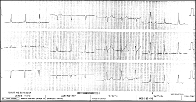
This is the EKG of a 24-year-old man who presents with chest pain. He has noted a low-grade fever over the past day or so. He usually enjoys good health.

Q. What do you think the diagnosis is?
A. The diagnosis is pericarditis. The diagnosis is suggested by the presence of the following changes:
- A depressed ST segment in aVR. This is virtually pathognomonic of pericarditis,
- Widespread elevation of the ST segment in leads I, II, III and aVF. There are no reciprocal changes.
- The ST segments are concave upwards. This used to be described as looking like 'Salvador Dali's moustache' but I guess that just dates me!
- There is evidence of PR depression, (Noted particularly in II and V4/5/6).
Q. What are the signs and symptoms of this condition and what might you expect to hear on cardiac auscultation?
A. The signs and symptoms in idiopathic or viral pericarditis include chest pain, fever, myalgias and a pericardial friction rub. The chest pain is classically described as worsening on lying down or on swallowing and relieved by sitting upright. It is believed that the pericardial friction rub is caused by friction between the inflamed pericardium and the surrounding tissues. The friction rub may be very localized but is often heard best at the lower left sternal border. It sounds creaky or scratchy and is best heard with the diaphragm of the stethoscope. Fever and myalgias are non-specific findings.
The treatment for pericarditis includes symptomatic therapy with non-steroidal agents. If the pain cannot be relieved, or if there is evidence of a pericardial effusion, the patient should be admitted. If there is any doubt about the diagnosis and a myocardial infarction is a possibility, admission should be considered. Some patients develop a chronic picture and these patients require steroid therapy.
Patient Number 3
This 46-year-old man presents to the ER stating "My heart seems to be going very fast. It woke me from sleep." He reports that this has happened once or twice in the past "but it always just settles down on its own". He is on no medications and has no other relevant past medical history. On physical exam, we find that he has a blood pressure of 124/84 and a pulse rate of about 200. This is his EKG.

Q. What does this EKG show?
A. This patient has very fast atrial fibrillation with aberrant atrio-ventricular conduction. The clues are found in the rhythm, which is irregular, and in the rhythm strip that shows the occasional normally conducted beat.

Q. What do you think the underlying problem might be?
A. This picture is seen classically in patients with Wolff Parkinson White (WPW) syndrome. WPW is an accessory pathway syndrome, where there is a connection from the atria to the ventricular myocardium. A rapid atrial fibrillation, like the one shown here, is seen in 10-30% of patients with WPW.

Q. How would you treat this in the ER?
A. Treatment of this rhythm would depend upon the clinical state of the patient. If the patient were awake, aware and hemodynamically stable, pharmacological management would be appropriate. If there is any sign of hemodynamic instability, the patient should be cardioverted emergently. In this patient's case, he was hemodynamically stable and so pharmacological therapy could be considered.
However, it is important to note a few potential problems. He is demonstrating so called 'antidromic' or aberrant conduction. In this situation, it is thought that the accessory pathway is being used as the anterograde limb and the AV node as the retrograde limb of the reentry circuit. The hallmark of this type of conduction is the broad QRS complex seen on this EKG, the irregularity and the presence of a delta wave.
Any wide complex, irregular arrhythmia at a rate of 250 or greater is highly suggestive of WPW with atrial fibrillation. When antidromic conduction is present, AV nodal blocking agents, for example beta-blockers, calcium channel blockers, adenosine and digoxin, are all contraindicated. This is because they may actually INCREASE the ventricular response because of enhanced conduction through the accessory pathway. Rapid ventricular rates can greatly increase the risk of ventricular fibrillation, and the rhythm can be intractable.
In these cases, the drug of choice is procainamide. The drug is administered I.V. at a rate of up to 50mg/min. The dose is usually in the range of 18-20 mgs/kg. Patients with congestive cardiac failure should receive a lower dose --12mgs/kg -- because they have reduced volume of distribution and decreased renal clearance. Many centers limit the total dose to 1000mgs but this may decrease the efficacy of this agent. The infusion should be stopped when cardioversion occurs, if hypotension intervenes or if the QRS widens to more than 50% above the pretreatment width.
This patient received a procainamide infusion, and cardioverted. His post cardioversion EKG is shown below and clearly demonstrates delta waves -- the slurred upstroke at the beginning of the QRS wave.

Patient Number 4
This 76-year-old female presented unconscious. There was no history available. She had not turned up at her regular morning bridge game and her friends were concerned. She was found on the floor of her room and brought to the emergency department. Her friends knew of no serious past medical history, though she was on a tablet for hypertension. She had seemed fine at breakfast and had just gone back to her room to 'freshen up.' On examination, she was unresponsive to pain. She had no obvious focal signs. Her pupils were midpoint and poorly reactive. This was her EKG.

Q. What is the diagnosis?
A. This patient has the 'giant T wave' syndrome. This unusual EKG appearance is seen related to myocardial ischemia apparently caused by increased left ventricular wall stress and changes in phasic coronary blood flow, from abnormalities in cerebral perfusion, particularly when associated with severe aortic regurgitation and, lastly, from coincidental neurological disease, particularly cerebral hemorrhage. This unfortunate lady had suffered a massive intracerebral bleed. She died later in hospital.

Patient Number 5
This 25-year-old man presented to the emergency room with 'palpitations'. This is his EKG.

Q. What is the diagnosis?
A. This patient has a narrow complex tachycardia, with a rate of about 150.
Q. What is the treatment?
A. The treatment for this man will depend upon his hemodynamic state. If he is hemodynamically unstable, he should have immediate cardioversion. One hundred joules is usually chosen as the initial dose and then increased until cardioversion occurs. Note that electrical cardioversion is rarely needed for rates below 150. If the patient is hemodynamicaly stable, the first 'treatment' suggested by the American Heart Association is a vagal maneuver. Examples of vagal maneuvers include carotid sinus massage (always listen for a bruit first!) and ice water facial immersion (contraindicated in patients with known ischaemic heart disease). If these efforts are unsuccessful, as they usually are, then pharmacological therapy is required.
The first line agent currently is adenosine. This drug has a very short half-life - in the order of about 10 seconds. It is eliminated by deamination in blood cells and in endothelial cells. Its onset of action is usually within 10 seconds and its short half-life is responsible for the treatment failures seen in about 25% of patients given the drug that initially cardiovert and then regress back to the tachycardia. The initial dose of adenosine is 6mgs and this is followed by up to two further doses of 12 mgs each, given 1 to 2 minutes apart. Any side effects of this medication are usually fleeting but include flushing, dyspnea, chest pressure, nausea and dizziness. Occasionally, patients may become hypotensive. Symptoms generally resolve without treatment but may be very unpleasant while they last.
If adenosine is not successful, second line agents, such as verapamil, should be considered. This drug is given as an I.V. bolus of 2.5 to 5 mgs initially. If this is tolerated, then a further bolus of 5 to 10 mgs can be given 10 to 15 minutes later. Some people use calcium chloride, 500 to 1000mgs (5 to 10 mls of a 10% solution) as prophylaxis against calcium channel blocker induced hypotension.
Should verapamil not be effective, other agents that may be considered include digoxin, beta-blockers and diltiazem. However, many would probably consider cardioversion at this point.
This patient responded to 12 mgs of adenosine.
Patient Number 6
This 56-year-old female presents at 0200 in the morning after having woken up with severe crushing central chest pain about 30 minutes earlier. She describes a vague ache in her chest with exertion over the past week or so. The ache had started to limit her activities but had always resolved completely with rest. This pain woke her and was associated with severe nausea and vomiting. The patient was diaphoretic and felt very anxious. Her past history was remarkable for hypertension managed with a diuretic. She smoked 20 cigarettes a day and had done so since the age of 15. Her father died of an infarct at age 42 and her mother had one heart attack at the age of 58. She has had no surgeries, experienced the menopause at age 52 and, until 2 weeks ago, considered herself in very good health. This was her presenting EKG.

Q. What abnormality is seen?
A. This patient has an acute inferior myocardial infarction. Inferior infarcts are characterized by ST segment elevation in leads II, II and aVF. It is important to note that up to 10% of patients with inferior myocardial infarction will present to the ED in complete heart block, two-thirds will develop complete block within 24 hours and the rest are likely to develop the problem within three days.
Q. What treatment would you recommend?
A. This patient needs to be considered for thrombolytic therapy. However, other agents are also important in the ER. The patient needs oxygen, nitroglycerin, initially sublingually, but then I.V. if there is no relief with sublingual, morphine in small incremental doses, and aspirin orally. The use of thrombolytics should be considered early, in an effort to reduce the 'door to needle' time - that time taken from the moment the patient arrives in the ED until they receive the thrombolytic. Other agents that should be considered acutely include beta-blockers and magnesium sulfate. Heparin infusion may also be required.
Q. What are the complete and the relative contraindications to thrombolysis?
A. The complete contraindications to thrombolysis include:
- Previous hemorrhagic stroke at any time, or any other stroke or cerebrovascular event within the last year
- Known intracranial neoplasm
- Active internal bleeding (EXCEPT menses)
- Suspected aortic dissection
The relative contraindications are:
- Severe, uncontrolled hypertension (BP > 180/110)
- Other intracranial pathology
- Current use of anticoagulants, or a known bleeding diathesis
- Trauma within past two weeks
- Prolonged CPR
- Major surgery within three weeks
- Venepunctures in non compressible sites
- Recent internal bleeding
- Pregnancy
- Active peptic ulcer
- History of chronic severe hypertension
- High chance of a left ventricular thrombus
- Acute pericarditis
- Subacute bacterial pericarditis
- Septic thrombophlebitis
- Diabetic hemorrhagic retinopathy
Q. Is she a candidate for this therapy?
A. This patient does not have any of the contraindications listed and is a candidate for thrombolysis.
This patient did receive thrombolysis, as well as a temporary pacemaker, and made a good recovery.
Diagnosis of an infarction is relatively straightforward when there are EKG changes like those illustrated. For cases that are less certain, there are many new cardiac markers now available. An earlier Cyberounds® presents a discussion of the management of myocardial infarctions.